
We recommend taking this quiz that will help you evaluate your knowledge before starting the tutorial.
The Formation of the heart tube is divided into several stages:
1- Establishment of the Cardiogenic Field
Cardiac progenitor cells (CPC) lie in the epiblast, lateral to the primitive streak where they migrate through. They are derived from intraembryonic mesoderm emerging from the cranial third of the primitive streak during early gastrulation.
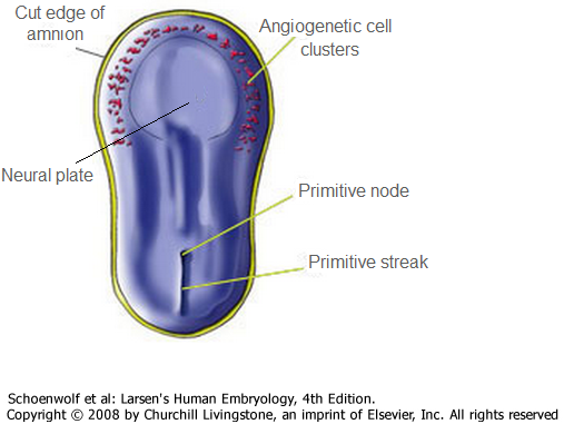
Figure 1
The cells proceed towards the cranium and position themselves anterior to the oropharyngeal membrane and neural folds as shown in fig.1 and fig.2.
The CPC will reside in the splanchnic layer of the lateral plate mesoderm, and later on they will be induced by the pharyngeal endoderm to form cardiac myoblasts.
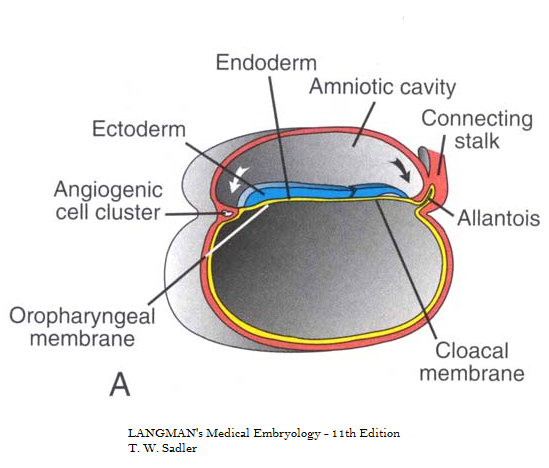
Figure 2
Blood island appear in this mesoderm, where they will form blood cells and vessels and with time islands unite to form the horseshoe-shaped tube surrounded by myoblast as shown in figure 1.
This region is called the cardiogenic field and the cavity over it later develops into the pericardial cavity as shown in figure 2.
2- Establishment of the heart Tube
Cephalocaudal folding Figure 3
Initially, the central portion of the cardiogenic area is anterior to the oropharyngeal membrane and the neural plate. During the process of body folding which is shown in figure 3, the cranial-most portion of the cardiac crescent swings ventrally and caudally to lie ventral to the newly forming foregut endoderm. |
Lateral folding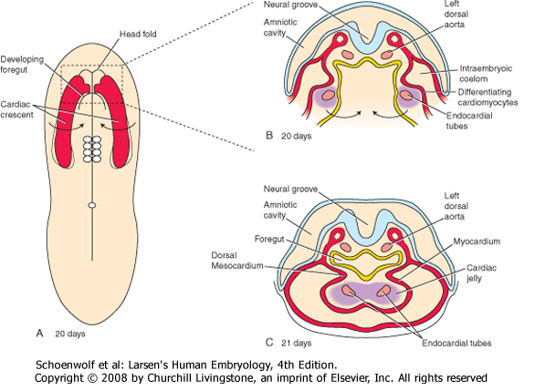 Figure 4
As the embryo folds cephalocaudally, it also folds laterally. As the lateral body folds move medially, they bring the right and left sides of the cardiac crescent together, and the two limbs of the crescent fuse in the midline, caudal to the head fold and ventral to the foregut. This fusion occurs at the site of the anterior intestinal portal and progresses in a cranial-to-caudal direction as the foregut tube lengthens. |
Simultaneously, the crescent part of the horseshoe-shaped area expands to form the future outflow tract and the ventricular regions.
By day 21, the primitive endocardial tube consists of an endothelium (i.e., endocardium) surrounded by a mass of splanchnic mesoderm containing cardiomyocytic progenitors that invest the fused endocardial tubes to form the myocardium, or heart muscle.
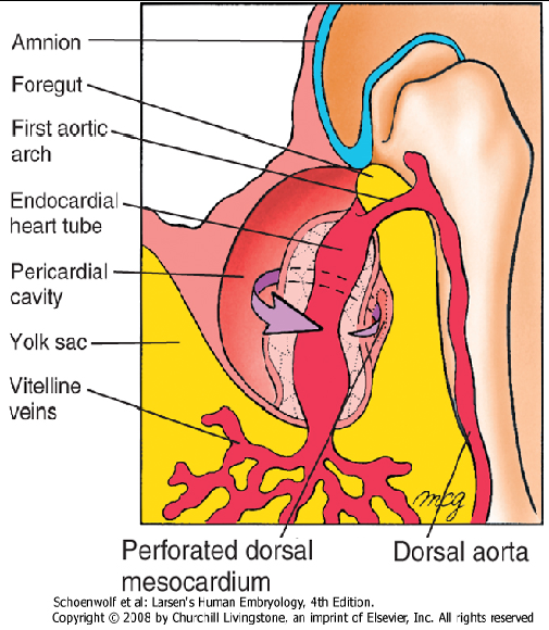
Figure 5
The primitive heart tube is initially suspended in the developing pericardial cavity by a dorsal mesocardium (dorsal mesentery of the heart) formed by splanchnic mesoderm located beneath the foregut.
This dorsal mesocardium promptly ruptures, leaving the heart suspended in the pericardial cavity by its attached vasculature. The region of the ruptured dorsal mesocardium becomes the transverse pericardial sinus within the pericardial sac of the definitive heart (fig. 5), a space separating the cardiac inflow and outflow vessels in adults.
Mesothelial cells on the surface of the septum transversum form the epipericardium near the sinus venous and migrate over the heart to form most of the epicardium.Thus the heart tube consists of three layers: the endocardium forming the internal endothelial lining of the heart , the myocardium forming the muscular wall and the epicardium or visceral pericardium covering the outside of the tube.
We recommend taking this quiz that will help you evaluate your knowledge after taking the tutorial.
Prepared by Joy Y. Balta
